|
|
|
||
| Home | About | Donate/Volunteer | Contact | Jobs| |
|
|
UCLA Researchers Map How Schizophrenia Engulfs Teen Brains | ||||||||||
|
UCLA Press Release - Monday, September 24, 2001 UCLA brain researchers using a powerful new technique have created the first images showing the devastating impact of schizophrenia on the brain. The findings, published in the Sept. 25 issue of the Proceedings of the National Academy of Sciences*, show how a dynamic wave of tissue loss engulfs the brains of schizophrenic patients in their teen-age years. The findings may have key diagnostic implications. Aided by a better understanding of how psychosis develops, researchers can detect aberrant loss early and treat patients as early as possible. Future medications might fight the rapid loss of brain tissue, and their effectiveness could be assessed using the imaging technique. "This is the first study to visualize how schizophrenia develops in the brain," said Paul Thompson, an assistant professor of neurology at the UCLA School of Medicine and the study's chief investigator. "Scientists have been perplexed about how schizophrenia progresses and whether there are any physical changes in the brain. We were stunned to see a spreading wave of tissue loss that began in a small region of the brain. It moved across the brain like a forest fire, destroying more tissue as the disease progressed." The scientists, at UCLA and the National Institute of Mental Health, employed magnetic resonance imaging (MRI) technology to scan a group of teenagers repeatedly as they developed schizophrenia. Using a new image analysis method that detects very fine changes in the brain, the scientists detected gray matter loss of more than 10 percent first in the parietal, or outer, regions of the brain; this loss spread to engulf the rest of the brain over five years. Patients with the worst brain tissue loss also had the worst symptoms, which included hallucinations, delusions, bizarre and psychotic thoughts, hearing voices, and depression. Schizophrenia affects an estimated 1 percent of Americans. Its causes are unknown, and the disease typically hits without warning in the late teens or 20s. Following is a selection of some of the pictures produced by the UCLA Laboratory of Neuro Imaging that show the impact of schizophrenia on the brain. For the full Press release and related press and journal articles go to: UCLA Researchers Map How Schizophrenia Engulfs Teen Brains
Mapping Brain Tissue Loss in Adolescents with Schizophrenia. This map reveals the 3-dimensional profile of gray matter loss in the brains of teenagers with early-onset schizophrenia, with a region of greatest loss in the temporal and frontal brain regions that control memory, hearing, motor functions, and attention. Using novel image analysis algorithms, dramatic reductions in the profiles of gray matter were detected, based on a database of 96 images from schizophrenic patients scanned repeatedly with MRI. The parallel extraction of anatomical models from all patients in the image database required 60 CPU hours, when running in parallel on an SGI RealityMonster with 32 internal CPUs. [Image by Paul Thompson, Christine Vidal, Judy Rapoport, and Arthur Toga].
Image 1. Frontal composite variability of normal and schizophrenia brains by gender
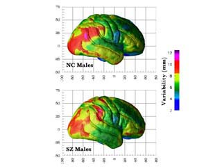
Image 2. Normal control vs. schizophrenia - Composite variability of 15 male subjects
Image 3. Volume of interest (VOI) superimposed over 3 orthagonal slices of the averaged schizophrenic brain This is a technique used to compare the volume of different areas of the normal control brain to a brain that has schizophrenia. Note the volume is simply a sphere with 60mm radius centered halfway between the midline decussations of the anterior and posterior commissures.
Image 4. Cortical surface variability maps. Variability maps of cortical surface and sulcal anatomy in normal controls (n = 28; 15 males) and schizophrenic patients (n =25; 15 males) showing the both hemispheres. The color bar indicates patterns of variability in each group as the root mean square magnitude of displacement vectors from each point in the surface meshes.
Image 5. Asymmetry maps Asymmetry maps were created in each group as defined by Sex and Diagnosis
(NC = normal controls, SZ = schizophrenic patients). Sulcal mesh averages
for each hemisphere were subtracted from a reflected version of the same
structure in the other hemisphere to create displacement vectors. These
maps
Image 6. 3D average surface representation and variability maps of the lateral ventricles Variability maps are similar in both groups with highest variability in the posterior horns (NC = normal controls, SZ = schizophrenic patients). Increases in LH ventricle length and volume were determined. The color bar encodes the root mean square magnitude of variability in millimeters.
Image 7. Displacements of the lateral ventricles and corpus callosum Displacement maps show the magnitude of displacement (mm) between schizophrenic patients and normal controls as represented by the color bar for the lateral ventricles and corpus callosum. A significant vertical displacement of the lateral ventricles in schizophrenic patients reflects a bilateral increase in ventricular volume, and corresponds to the displacement of the corpus callosum.
Image 8. Cortical surface variability maps. Variability maps viewed from the front showing cortical surface variability
in the four groups defined by sex and diagnosis.
Additional pictures and images of brains with schizophrenia can be viewed at http://www.loni.ucla.edu/ picture of schizophrenia, picture and schizophrenia, image and schizophrenia, brain and schizophrenia, pictures, photos, photographs, image, pictures picture of schizophrenia, picture and schizophrenia, image and schizophrenia, brain and schizophrenia, pictures, photos, photographs, image, picturespicture of schizophrenia, picture and schizophrenia, image and schizophrenia, brain and schizophrenia, pictures, photos, photographs, image, pictures
|
Advertisement
|